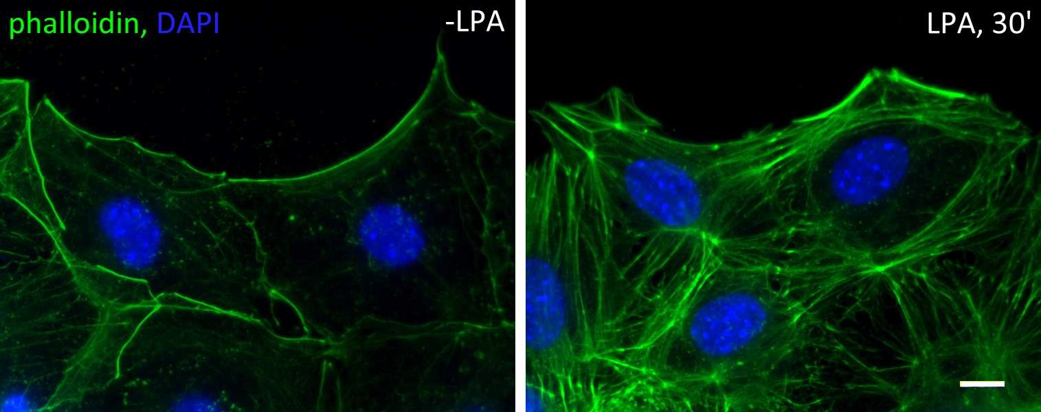
Serum-starved fibroblasts lose most of their filamentous actin structures. Upon stimulation of serum (or serum factor lysophosphatidic acid), rho and cdc42 GTPases are activated. Rho activation leads to the formation of stress fibers, while cdc42 activation leads to the establishment of cell polarity. Cdc42 activates myotonic dystrophy kinase-related cdc42-binding kinase (MRCK) to phosphorylate the regulatory light chain of myosin II, allowing the formation of actin cables at the cell cortex.
In cells at the wound edge of a monolayer, these cables are formed mostly at the leading edge, i.e. the wound edge. These cables are highly dynamic. They form constantly and are driven by mysoin II to move retrogradely. Two myosins II are essential – myosin IIA is the engine and drive the movement, and myosin IIB is the steering wheel that controls the direction. In the absences of myosin IIA no actin cables can form or move; whereas in the absences of myosin IIB the actin cable flow is chaotic and move in random directions. Below is a movie of a lysophosphatidic acid-activated cells, red is an actin probe mCherry-LifeAct and green is the nucleus.