Particle Analyzer is a free, stand-alone Windows program. Current version is v0.6. It is provided “as-is”, without any express or implied warranty.
Particle Analyzer is a newer version of Quantify Colocalization. By using local background subtraction and other mathematical morphology operations, the program is able to detect hundreds of particles of various sizes and shapes almost instantly. After opening the images, the user draws the boundaries of the cells, and the program will detect particles in the cell and calculate their positions, sizes, signals from each channel, distances to the membrane and cell centroid.
Feature
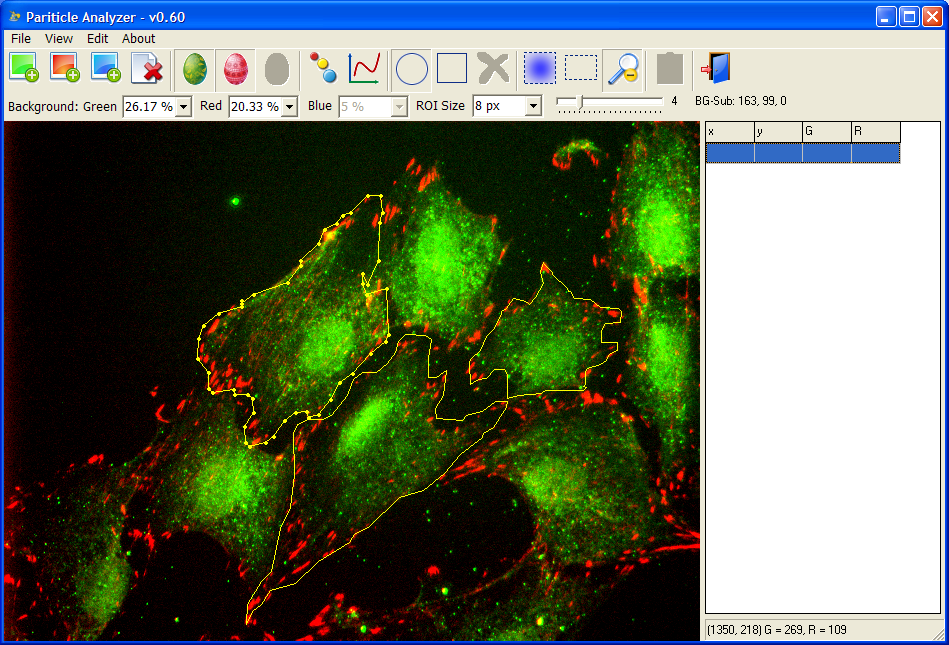
- Draw cell boundary directly on the images with a mouse. Cell boundary can be freely modified afterwards.
- All particles within a cell boundary will be analyzed and exported.
- Result of particle detection can be visualized.
- Some settings of particle detection can be adjusted.
- Has intuitive built-in background subtraction. Simple click the set background icon (5th icon from the right) and select a rectangle.
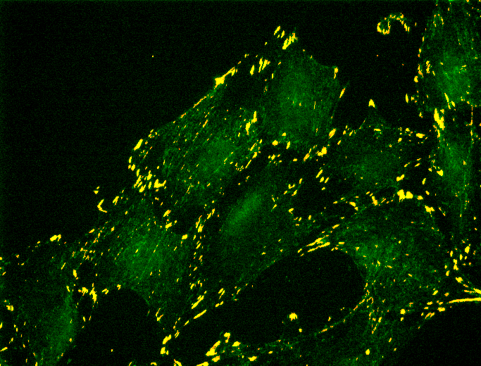
Particle Analyzer detects particles pretty well. Here is a merged image of focal adhesion (Green, raw image) and detected particles (Red). Note that there is no false-positive (pure red) particles and most of the diffusive background (pure green) signals are excluded.
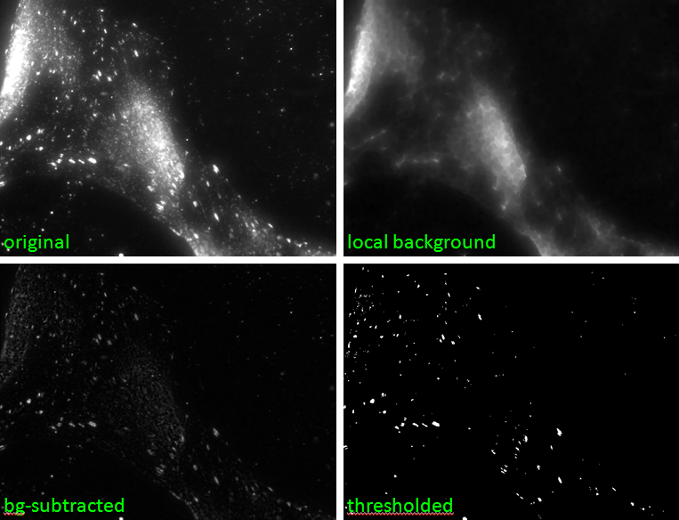
Particle Analyzer is also good at dealing with large noise. The following is an image with strong nuclear noise. They are removed by local background subtraction. Several internal steps are shown below.
How to use
To try using Particle Analyzer, download the program with sample images and extract the files to a folder. The files are CountSignal.exe and two images files: Green.tif and Red.tif.
- Double click to run CountSignal.exe.
- Drag and drop Green.tif.
- Drag and drop Red.tif. Note that, for now, the program always uses the second channel for particle detection. So the sequence of channel loading is important.
- When holding the Ctrl key, use the mouse to draw a cell boundary.
- Double click to end boundary drawing.
- Repeat for all cells of interest.
- Copy results and paste to Excel
Particle Analyzer will be updated to support stack images and images with more than three channels in the future. Cell boundary and nucleus detection, particle detection with other channels, user-defined parameters will also be supported. Particle tracking in movies is also planned but will take longer.