Drawing of nuclear transport and the NPC

Chen M, Ma Y, Chang W. SARS-CoV-2 and the Nucleus. International Journal of Biological Sciences, 2022 Jul; 18(12):4731-4743.
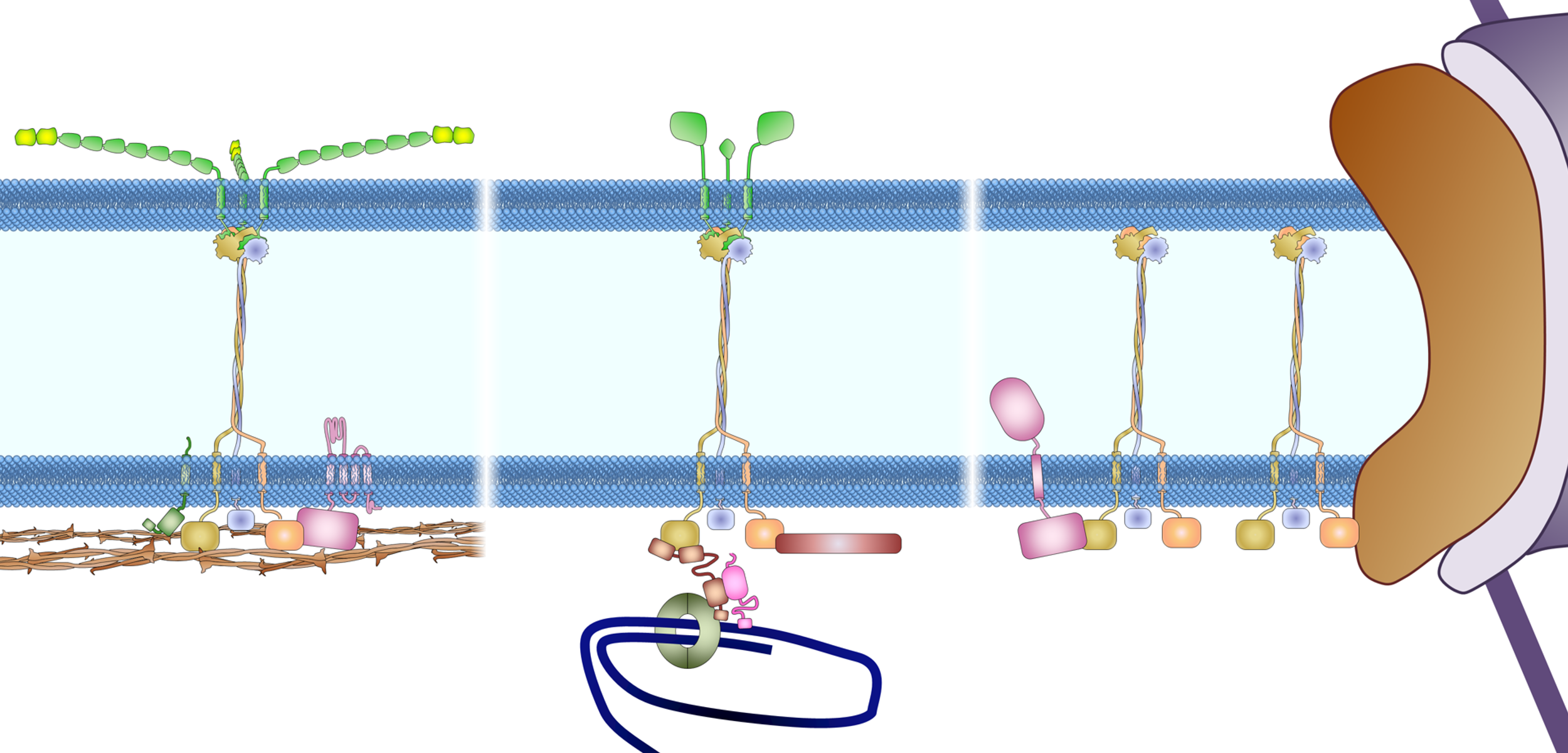
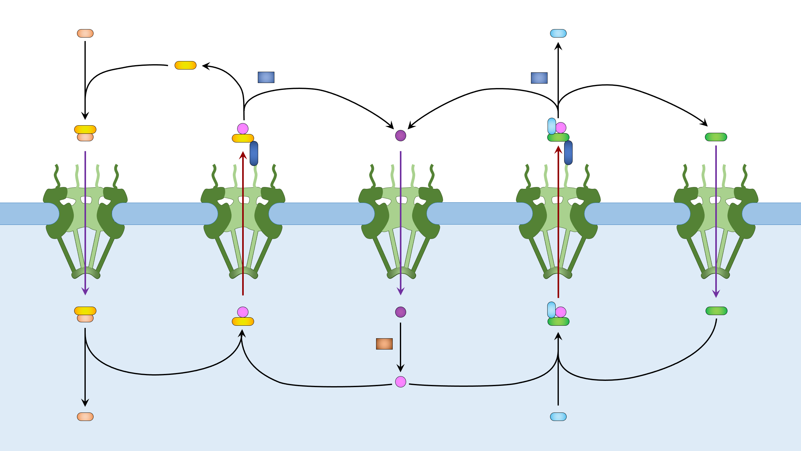
Figure 1. Nuclear import and export. Left: Cargo to be imported binds to cytoplasmic importin and enters the nucleus, where it is released from importin by Ran-GTP. The Ran-GTP/importin complex moves to the cytoplasm and binds RanBP. Ran-GAP activates the GTPase activity of Ran to convert Ran-GTP to Ran-GDP and release importin. Ran-GDP enters the nucleus and is recharged by a Ran-GEF. Right: Cargo to be exported binds to exportin and Ran-GTP in the nucleus. The complex moves to the cytoplasm where Ran-GTP is converted to Ran-GDP, leading to the disassembly of the complex. Both Ran-GDP and exportin then enter the nucleus for another round of nuclear export.
Nuclear import and export, with lables (2560 × 1440 px).
Nuclear import and export, without lables (2560 × 1440 px).
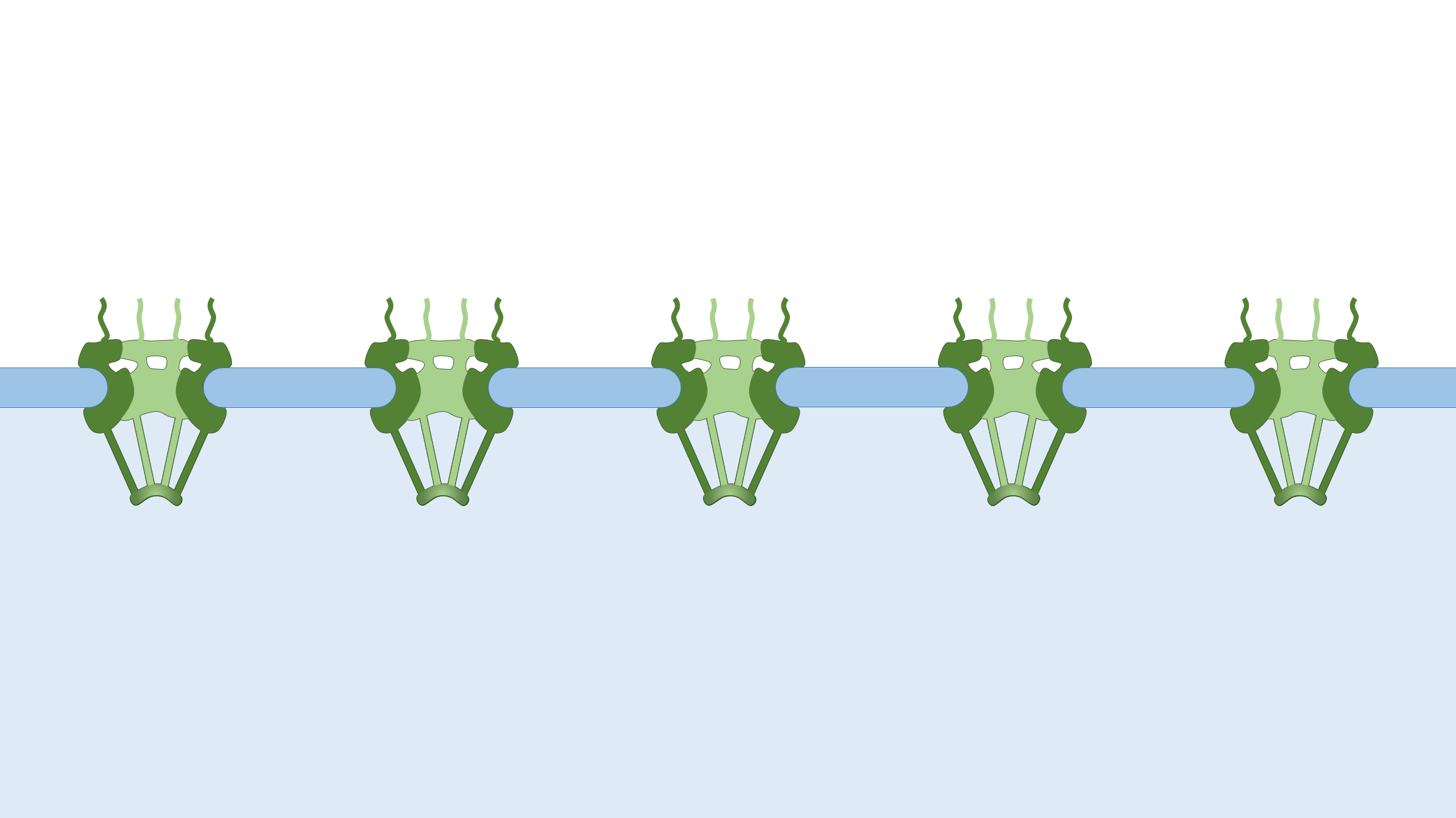
The nuclear pore complexes (2560 × 1440 px).

Drawing of the nuclear envelope

Li Y, Chen M, Chang W. Roles of the nucleus in leukocyte migration. Journal of Leukocyte Biology, 2022 Oct; 112(4):771-783.
Figure 2. The LINC complexes connect the cytoskeleton to the nuclear lamina and chromatin in somatic cells. The LINC complexes are composed of nesprins on the ONM and SUN proteins on the INM. The cytoplasmic domains of nesprins interact with the cytoskeleton. The giant isoforms of nesprin-1 and -2 contain actin binding domains and interact with actin filaments. Such an interaction is regulated by emerin through its interaction with myosin II and is strengthened by fascin and formin proteins FHOD1 and FHOD3. Nesprin-1α interacts with AKAP6 and AKAP9 to facilitate microtubule nucleation. Both the long and short isoforms of nesprin-1 and −2 contain a kinesin-1- and dynein-binding region and associate with microtubules through the motors. Nesprin-4, which may not be expressed in leukocytes, can also bind to kinesin-1. Nesprin-3 binds to plectin (and the related BPAG1 and MCAF), which can interact with both microtubules and intermediate filaments. In the perinuclear space, trimeric SUN proteins bind to nesprins and torsin A. In the nucleoplasm, SUN proteins bind to lamins for anchorage and interact with Samp1 and emerin. Many of these proteins, like lamin A, lamins B (through LBR), SUN proteins (through an unknown adaptor), and LEM domain proteins (Man1, LAP2, emerin, and LEMD2, through BAF and HDAC3) directly or indirectly bind to chromatin.
The nuclear envelope, with lables (2560 × 1320 px).
The nuclear envelope, without lables (2560 × 1320 px).

Drawing of the anchorage of the LINC complex

Chang W, Worman HJ, Gundersen GG. Accessorizing and anchoring the LINC complex for multifunctionality. Journal of Cell Biology, 2015 Jan 5; 208(1):11-22.
Figure 3. Anchoring the LINC complex. Mammalian SUN proteins are anchored to the inner nuclear membrane through at least three different mechanisms. (A) The nucleoplasmic tail of SUN2 binds to lamin A and anchors the LINC complex to the nuclear lamina in somatic cells. Samp1 and emerin are required to strengthen this anchorage during nuclear movement, presumably to resist the high mechanical force. For clarity, nesprin-2G is shown as a shorter protein without all of its 56 SRs. INM, inner nuclear membrane; ONM, outer nuclear membrane. (B) The nucleoplasmic tail of SUN1 binds TERB1 and anchors the LINC complex to chromosomes through telomere binding proteins (TRF1 and cohesion) in meiotic cells. Lamin C2 also associates with this complex, probably through SUN1 binding. (C) Nucleoplasmic tails of SUN proteins shown binding to nuclear pores (SUN1) and a hypothetical protein as possible alternative anchors for the LINC complex in somatic cells. As described in the text, the localization of SUN1 and SUN2 in the nuclear membrane is only slightly affected in somatic cells lacking all lamins, which indicates the presence of additional anchoring factors.
Anchoring the LINC complex, with lables (2500 × 1200 px).
Anchoring the LINC complex, without lables (2500 × 1200 px).

Drawing of the LINC complex

Chang W, Worman HJ, Gundersen GG. Accessorizing and anchoring the LINC complex for multifunctionality. Journal of Cell Biology, 2015 Jan 5; 208(1):11-22.
Figure 1. The LINC complex bridges the cytoskeleton and nucleoskeleton. The LINC complex is composed of KASH proteins in the outer nuclear membrane and SUN proteins in the inner nuclear membrane. The lumenal region of SUN proteins forms a triple helical coiled-coil, allowing trimerization of their SUN domains. The hydrophobic groove between neighboring SUN domains is required for the KASH peptide to bind, and this interaction is further strengthened by a KASH-lid of the SUN domain. The cytoplasmic extensions of KASH proteins vary in size and interact with different cytoskeletal elements. The nucleoplasmic domains of SUN proteins anchor the LINC complex to the nucleoskeleton, through its interaction with nuclear lamina, as well as chromosome-binding proteins and probably other anchoring proteins. INM, inner nuclear membrane; ONM, outer nuclear membrane.
A LINC complex with actin filaments, with lables (1600 × 1600 px).
A LINC complex with actin filaments, without lables (1600 × 1600 px).
A LINC complex with both microtubule and actin filament, with lables (1600 × 1600 px).
A LINC complex with both microtubule and actin filament, without lables (1600 × 1600 px).