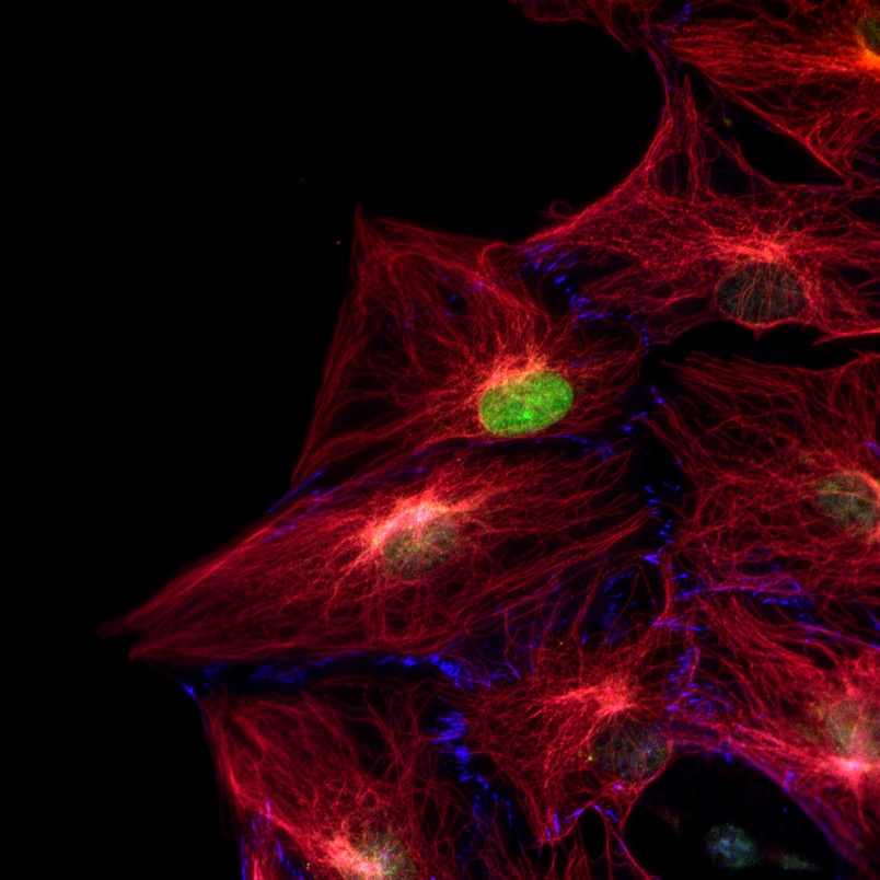
First data in our lab is a fluorescent image taken on a Nikon Ti microscope. NIH 3T3 fibroblasts were microinjected with plasmid DNA encoding GFP-prelamin AL647R for 1 hour and cell polarization was induced by 10 μM lysophosphatidic acid. Two hours later cells were fixed and stained for tubulins (red) and vinculin (blue).
